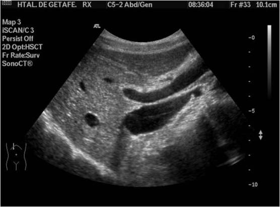
La Vesícula es una de las estructuras más importantes dentro del estudio de Abdomen porque gran cantidad de estudios se van a realizar bajo la sospecha clínica de patologías que tienen que ver con esta estructura. Si bien la Vesícula es una estructura adosada al Hígado, se engloba en el estudio hepático por motivos obvios.
Conocer su anatomía y la de sus conductos nos hará comprender que es una estructura cuya participación en la digestión y en los procesos que se llevan a cabo en el abdomen superior están íntimamente relacionados e interconectados, sobre todo con el Páncreas.
La vesícula es un órgano oval en forma de saco que se divide en: Infundíbulo, Cuerpo y Fundus.
Los conductos biliares se dividen en porción intrahepática y extrahepática.
Los conductos intrahepáticos discurren en paralelo a las venas porta
La porción extrahepática incluye el conducto hepático común, el colédoco y una porción de los conductos centrales dcho e izdo.
Me parece muy importante que conozcas este detalle de cómo «convive» la Vesícula y sus Conductos con otras estructuras colindantes como la Arteria Hepática, y la Vena Porta en el hilio hepático, porque ecográficamente son estructuras milimétricas, en ocasiones virtualmente invisibles, pero vitales.
El Colédoco es una estructura muy importante, también se la conoce como vía biliar extrahepática y discurre paralela y anterior (ecográficamente hablando) a la Vena Porta Extrahepática en el hilio hepático.El Colédoco es una estructura que en ocasiones no vemos en ecografía ya que se estima que por cada 10 años que cumple un ser humano, la estructura crece 1 mm, por tanto es normal que en edades menores de la treintena, no lo veamos.
La Porta es nuestra guía para controlar al Colédoco y junto a ellos, muy pequeña, la Arteria Hepática, desconocida, pero que se visualizan siempre juntos y hay que saber distinguir, las estructuras vasculares, Porta y Arteria Hepática del Colédoco, ¿pero cómo?, muy fácil, con el Doppler Color, Vena y Arteria tienen flujo, el Colédoco, no.
Para el estudio de la Vesícula es incondicional las ayunas de la/el paciente.
El estudio ecográfico de la vesícula se tiene que realizar haciendo un corte longitudinal (negro) y transverso (verde) de la estructura, como en la imagen siguiente, el colédoco por su parte, debe verse, si se viera, paralelo en un corto espacio, a la Porta extrahepática, como dos estructuras alargadas.
The Vesicle is one of the most important structures in the study of Abdomen because a large number of studies are going to be carried out under the clinical suspicion of pathologies that have to do with this structure. Although the Vesicle is a structure attached to the Liver, it is included in the liver study for obvious reasons. Knowing its anatomy and that of its ducts will make us understand that it is a structure whose participation in digestion and in the processes that are carried out in the upper abdomen are closely related and interconnected, especially with the Pancreas. The vesicle is an oval organ in the form of a sac that is divided into: Infundibulum, Body and Fundus. The bile ducts are divided into intrahepatic and extrahepatic portions. The intrahepatic ducts run parallel to the portal veins The extrahepatic portion includes the common hepatic duct, the common bile duct, and a portion of the right and left central ducts. It seems very important that you know this detail of how the Vesicle and its Conduits «coexist» with other adjacent structures such as the Hepatic Artery, and the Porta Vena in the hepatic hilum, because ultrasound are millimeter structures, sometimes virtually invisible, but vital. The common bile duct is a very important structure, it is also known as extrahepatic bile duct and runs parallel and anterior (echographically speaking) to the extrahepatic portal vein in the hepatic hilum. The common bile duct is a structure that we sometimes do not see in ultrasound since it is It estimates that for every 10 years that a human being fulfills, the structure grows 1 mm, so it is normal that in ages under thirty, we do not see it. The Porta is our guide to control the common bile duct and, very small, the hepatic artery, unknown, but always visualized together and we must know how to distinguish the vascular structures, Porta and Hepatic Artery of the common bile, but how? , very easy, with the Color Doppler, Vein and Artery have flow, the Choledochus, no. For the study of the Vesicle, the fasting of the patient is unconditional. The ultrasound study of the gallbladder must be done by making a longitudinal (black) and transverse (green) section of the structure, as in the following image, the bile duct, on the other hand, should be seen, if seen, parallel in a short space , to the extrahepatic Porta, as two elongated structures.

Para visualizar la Vesícula pediremos a la/el paciente que esté en inspiración forzada, realizaremos un corte sobre el eje largo de la vesícula (negro) y otro sobre su eje corto (verde), longitudinal y transverso respectivamente.
Ecográficamente la Vesícula tiene un aspecto sacular y anecoico de paredes finas.Puede tener varias presentaciones anatómicas todas ellas normales, es decir, su forma, aunque sacular y normal, no siempre es igual, varía entre pacientes. Obsérvese el pictograma.
To visualize the Vesicle we will ask the patient to be in forced inspiration, we will make a cut on the long axis of the vesicle (black) and another on its short axis (green), longitudinal and transverse respectively. Ecographically the Vesicle has a saccular and anechoic aspect with thin walls. It can have several anatomical presentations, all of them normal, that is, its shape, although saccular and normal, is not always the same, varies between patients. Observe the pictogram.

El Colédoco es por su parte, ecográficamente, anecoico y alargado, pero serpenteante en ocasiones.
The Choledochus is, on the other hand, echographically, anechoic and elongated, but sometimes serpentine

El Conducto hepático común es anatómicamente hablando un gran desconocido para los Técnicos, pero acompaña a la Porta Intrahepática, no suele visualizarse ecográficamente, y si lo hace se identifica como una doble vía y está en relación con procesos patológicos hepáticos.
Para el estudio de la Vía Biliar Extrahepática o Colédoco realizaremos un corte para sagital al eje largo de la Línea Alba (negro) y para ver la Vía Biliar Intrahepática, lo haremos axial o para axial al cuerpo (rojo), siempre en función de la estructuras como la imagen que ves a continuación y recomiendo realizar este estudio y el de la Vesícula tanto en decúbito supino como en decúbito lateral izquierdo y escoger las mejores visualizaciones para realizar las fotos.
The common hepatic duct is anatomically speaking a great unknown to the technicians, but it accompanies the intrahepatic portal, it is not usually visualized ecographically, and if it does it is identified as a double pathway and is related to liver pathological processes. For the study of the Extrahepatic Biliary or Copedopathic Way we will make a sagittal cut to the long axis of the Alba Line (black) and to see the Intrahepatic Biliary Path, we will do it axially or axially to the body (red), always depending on the structures like the image that you see below and I recommend doing this study and that of the Vesicle both in the supine position and in the left lateral decubitus and choose the best visualizations to take the photos


En ambas imágenes superiores, tanto vía biliar intrahepática como la extrahepática acompañarían a ambas porciones de la Vena Porta. En la siguiente imagen vemos Porta Intrahepática con la Vía biliar intrahepática acompañando al vaso, anterior y ambas imágenes paralelas de aspecto ecográfico normal y anecoico, claro está.
In both superior images, both intrahepatic and extrahepatic biliary tract would accompany both portions of the portal vein. In the following image we see Porta Intrahepatic with intrahepatic biliary path accompanying the vessel, anterior and both parallel images of normal and anechoic echographic appearance, of course.

Ahora con textos:

En la siguiente imagen, Vesícula, Vía Biliar Extrahepática y pequeña porción de Vena Porta:

Ahora, con textos…

Cuando hablemos de la patología biliar conoceremos mucho más y veremos más de la imágenes, pero hoy solo importaba la técnica y el aspecto ecográfico.
Post de difícil comprensión por la gran cantidad de información.
Vital la comprensión de la anatomía y sus interrelaciones.
Para el protocolo, nos guardamos la foto de ambas Venas Portas y eventual visualización de las vías biliares correspondientes y por supuesto la foto de la Vesícula Biliar.
Seguimos…
Muy buena y sencilla explicación .
Me gustaMe gusta
Muchas gracias.Gracias por visitar mi Blog.
Me gustaMe gusta
Buenas noches, tengo una pregunta, si al momento de evaluar el paciente valoró la vesícula y confirmó que tiene una colelitiasis, pero no veo el colédoco dilatado, en que tiempo se podría producir una dilatación de vías biliares, producto de la colelitiasis ??
Me gustaMe gusta
Pues no lo sé, cuando una piedra sale de la vesícula y queda en el colédoco se verá dilatado, pero no es una causa única.Espero haber podido contestar a su pregunta.
Me gustaMe gusta
Hola, soy médica de familia y me estoy iniciando en la ecografía. Muchas gracias por esta labor altruista de docencia y por este magnífico blog
Me gustaMe gusta
María José, no sabe lo bien que me vienen estas palabras amables. Son la única gasolina que me impulsa a seguir. Gracias.
Si puedo ayudarla en cualquier cosa no dude en contactar a través de los canales de Blog de Twitter e instagram o si prefiere, por correo electrónico.
Mil Gracias.
Me gustaMe gusta
Hola, buenas tardes soy médico ultrasonografista, y en su página he encontrado muchos tips para mi consulta, pero hay algo que me gustaria preguntarle, existe alguna bibliografia que hable sobre el uso de focos adecuadamente según cada valoración, por ejemplo cuantos en tiroides, cuantos en mama, cuantos para valoración de higado, y así?, mi duda es porque en mi trabajo nos piden protocolos rígidos sobre su uso y no creo estar a favor de eso, ya que yo soy de usarlo de acuerdo a las necesidades, pero ellos nos piden 2 focos para todo, no se me hace correcto, igual me gustaría saber si existe alguna bibliografía de focos.
Me despido agradeciendo de antemano su tan relevante información.
Me gustaMe gusta
HOLA SOY MEDICO ULTRASONOGRAFISTA Y SI HAY BIBLIOGRAFIA SOBRE LO QUE DEBEMOS DE REPORTAR EN CADA ORGANO, LO PUEDE ENCONTAR EN LA PAGINA DE SCRIBD
Me gustaMe gusta