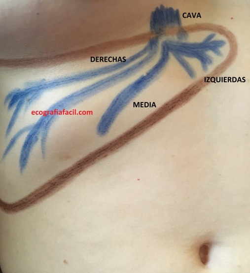
Las venas Suprahepáticas son vasos que van a parar a la Cava llevando sangre «sucia». Es primordial conocer su Anatomía. básicamente tenemos que saber en que se dividen en 3 bloques, Izquierda, Intermedia o media y Derecha, como tres bloques principales, luego, como si de ramas de un árbol se tratase, se subdividen según cada anatomía, así:
The Suprahepatic veins are vessels that go to the Cava carrying «dirty» blood. It is essential to know your Anatomy. Basically we have to know in which they are divided into 3 blocks, Left, Intermediate or Middle and Right, as three main blocks, then, as if they were branches of a tree, they are subdivided according to each anatomy, like this:
Si observas en la imagen, el Hígado está protegido por la parrilla costal, las Suprahepáticas son proximales y por tanto para poder estudiarlas tendremos que apoyarnos en el reborde costal inferior y angularnos para poder estudiarlas, ya que justo encima de ellas es imposible virtualmente debido a las interfases provocadas por el hueso de las costillas, es decir, nos tenemos que colocar así:
If you look at the image, the Liver is protected by the rib cage, Suprahepatic proxies are proximal and therefore to study them we will have to rely on the inferior costal rim and angles to be able to study them, since just above them is virtually impossible due to the interfaces caused by the bone of the ribs, that is, we have to place it like this:
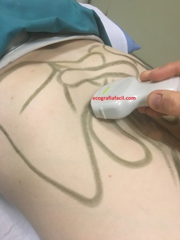
Esta imagen es el secreto de la imagen de hoy y cómo conseguirla. Localiza el reborde costal, tócalo, coloca el transductor paralelo a él, angula como si pusieses el transductor casi paralelo a la piel del paciente y busca las Suprahepáticas sabiendo su disposición anatómica, el resultado, este:
This image is the secret of today’s image and how to get it. Locate the costal flange, touch it, place the transducer parallel to it, angle as if you placed the transducer almost parallel to the skin of the patient and look for the Suprahepatic knowing its anatomical disposition, the result, this:

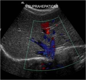
Respecto de la imagen anatómica puede parecer que la imagen está al revés, pero piensa que donde apoyamos la sonda es la zona superficial de la imagen ecográfica y la parte más profunda de la imagen correspondería con la anatomía que nos interesa estudiar.
Vuelvo a recordar, aquí los cortes son en extricta referencia a la estructura estudiada, no son cortes puros a los ejes del abdomen.
El aspecto ecográfico son de estructuras anecoicas, sin un marcado perfil hiperecogénico al contrario que la Porta, alargadas e intrahepáticas que se subdividen en ramas partiendo de tres ejes principales.
Regarding the anatomical image it may seem that the image is upside down, but think that where we support the probe is the surface area of the ultrasound image and the deepest part of the image would correspond to the anatomy that we are interested in studying. I remember again, here the cuts are in strict reference to the structure studied, they are not pure cuts to the axes of the abdomen. The echographic aspect is of anechoic structures, without a marked hyperechogenic profile unlike the Porta, elongated and intrahepatic that are subdivided into branches starting from three main axes.
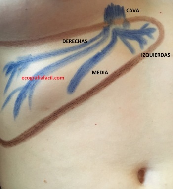

Estas dos imágenes son la base del post de hoy junto con el posicionamiento de la sonda…
Ahora bien, recomiendo enfervorecidamente el estudio de cada una de las suprahepáticas de modo individual, y empezamos por la Suprahepática Izquierda. Para ello colocaremos la sonda como para el estudio del post anterior y la angularemos en función de la anatomía de la Suprahepática Izquierda, es decir, así:
These two images are the basis of today’s post along with the positioning of the probe … Now, I strongly recommend the study of each one of the suprahepatic ones in an individual way, and we start with the Left Suprahepatic. To do this we will place the probe as for the study of the previous post and we will angle it according to the anatomy of the Left Suprahepatic, that is, like this:

y tenemos que obtener este resultado:
and we have to obtain this result:

Obsérvese en la imagen el pictograma.
Para la media y la suprahepática derecha se puede usar la colocación base, la primera que he contado e ir buscando cada una de forma individualizadamente hasta conseguir desplegarlas y lograr una imagen como esta:
Observe in the image the pictogram. For the mean and the right suprahepatic, the base placement can be used, the first one that I have counted and go searching each one individually to get them to unfold and achieve an image like this:

Cuando nos enfrentamos a anatomías complicadas, hígados subcostales, mucho gás abdominal, etc, es interesante pedirle a la/el paciente que adopte la posición en decúbito lateral izquierdo para obtener los resultados más óptimos posibles, colocando nuestra sonda igual que si estuviese en decúbito supino. Y por cierto, en ocasiones podemos ver estos vasos mejor en apnea que en inspiración, pero siempre hay que probar ambas opciones.
Estos vasos pueden verse excesivamente grandes cuando hay patologías congestivas cardiacas y pueden llegar a llamar la atención por lo «llamativamente grandes que se ven», ojo, que es un signo de patología en este caso de posible origen cardiaco, pero corresponde al Radiólogo la valoración, eso sí, nosotros siempre atentos a cosas «no normales».
Muy interesante estos vasos, cuya circulación sanguínea aleja la sangre de la sonda y por tanto si aplicamos el modo de trabajo doppler color, veremos que su imagen es azul..
When faced with complicated anatomies, subcostal livers, much abdominal fat, etc., it is interesting to ask the patient to adopt the position in the left lateral decubitus to obtain the best possible results, placing our probe as if in the supine position. . And by the way, sometimes we can see these glasses better in apnea than in inspiration, but you always have to try both options. These vessels can be excessively large when there are congestive cardiac pathologies and can get attention because of the «strikingly large that are seen», eye, which is a sign of pathology in this case of possible cardiac origin, but the radiologist corresponds to the assessment , yes, we are always attentive to «not normal» things. Very interesting these vessels, whose blood circulation moves the blood away from the probe and therefore if we apply the Doppler color working mode, we will see that its image is blue ..

Excelente presentación la primera vez que veo tu pag
Me gustaMe gusta
Muchísimas gracias, espero que le sea útil y si es así, que lo difunda, le animo a suscribirse y a compartir.Gracias de corazón.
Me gustaMe gusta
Muy bueno los temas expuestos en su página de verdad. No encontré otra que sea muy didáctico y comprensible como los temas q están en su página. Espero siga a ese ritmo con nuevos temas. Y gracias. Q m ayudó bastante en mi trabajo.
Me gustaMe gusta
Muchas gracias a usted por su comentario, le pido que difunda y comparta el Blog para que llegue a más gente.
Me gustaMe gusta
Muchas gracias! Nos facilitas la vida!!
Me gustaMe gusta
jajajajajaja, me encanta oir eso¡¡¡
Me gustaMe gusta